New Patients
Existing Patients
New Patients
Existing Patients
New Patients
Existing Patients
New Patients
Existing Patients
At Myers Pediatric Dentistry & Orthodontics, we combine a child-focused approach with modern diagnostic tools to deliver confident, evidence-based care. One of the technologies we rely on is cone-beam computed tomography (CBCT), a 3D imaging method that captures detailed views of teeth, jaws, and surrounding anatomy that traditional two-dimensional X-rays cannot show.
CBCT helps our team make more informed decisions while keeping patient comfort and safety at the forefront. The images produced are precise enough to guide treatment planning yet obtained quickly and with focused exposure, which is particularly important when treating young patients.
Cone-beam computed tomography (CBCT) creates three-dimensional images by rotating a cone-shaped X-ray beam around the patient. The resulting dataset can be reconstructed into multiple views — axial, coronal, sagittal, and three-dimensional renderings — allowing clinicians to evaluate anatomy from every angle. This gives a fuller picture than conventional bitewing or panoramic X-rays.
For pediatric patients, the ability to see structures in three dimensions is especially useful. CBCT reveals the position of unerupted or impacted teeth, the relationship between developing roots, and the shape of jawbones and sinuses. Those details can be pivotal when planning orthodontic treatment or assessing developmental concerns.
Importantly, CBCT is not a replacement for routine 2D X-rays in every situation. Our team chooses imaging intentionally — balancing diagnostic benefit with the principle of keeping exposure as low as reasonably achievable — reserving CBCT for cases where the added information will meaningfully influence care.
When complex anatomy or unclear findings are present, CBCT can change the clinical picture. It helps detect hidden pathology, define the precise location of an impacted tooth, clarify the cause of facial pain, and assess bone volume and structure when surgical planning is required. These insights reduce guesswork and support more predictable outcomes.
For orthodontic care, CBCT aids in evaluating jaw relationships and airway space, assisting providers in developing treatment strategies that account for growth and function. The three-dimensional perspective improves appliance positioning and monitoring, helping clinicians tailor interventions to each child’s unique anatomy.
In trauma or emergency scenarios, CBCT can reveal fractures and displaced teeth that may not be obvious on standard films. That fuller diagnostic view speeds appropriate clinical decisions and supports focused, conservative treatment when possible.
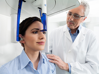
A CBCT scan at our office is designed to be quick and comfortable. Patients are positioned either sitting or standing, and the scanner rotates around the head for a matter of seconds. Because the scan is brief and noninvasive, most children tolerate it well without the need for sedation or lengthy preparation.
The procedure feels similar to an ordinary X-ray appointment. Technicians use stabilizing aids and clear instructions to reduce movement and capture the best images. Radiation protection measures are in place, and the field of view is tailored to the area of interest to limit exposure to surrounding tissues.
After the scan, the images are reviewed by our clinicians using advanced viewing software that allows cross-sections, measurements, and three-dimensional manipulation. This visual information makes it easier to explain findings to parents and to plan next steps with clarity.
CBCT differs from traditional medical CT in that it uses a focused cone-shaped beam and a single rotation to produce volumetric images. That design enables excellent spatial resolution for dental and maxillofacial structures while often using a lower radiation dose than medical CT scans. Adjustable settings allow clinicians to prioritize resolution or minimize exposure depending on the clinical need.
Modern CBCT systems also provide selectable fields of view. For instance, when only a few teeth or a limited region require evaluation, the scan can be confined to that area rather than imaging the entire craniofacial complex. This targeted approach reduces unnecessary exposure while still yielding the high-detail images needed for precise analysis.
Image-processing tools refine raw data into clinically useful views: thin-slice cross-sections, panoramic reconstructions, and lifelike 3D renderings. These views make it possible to measure distances, evaluate root morphology, identify anatomic landmarks, and simulate treatment outcomes with greater confidence than two-dimensional images alone.
CBCT images serve as a common visual language among dental and medical providers. When specialty collaboration is needed — for orthodontics, oral surgery, or airway assessment — three-dimensional scans make it easier to discuss options, share precise measurements, and align on a coordinated plan of care for the child.
Digital CBCT datasets integrate smoothly into treatment workflows, enabling the team to fabricate guides, plan minimally invasive procedures, and monitor growth or healing over time. The result is a more streamlined process and clearer decisions based on objective anatomic information.
Clinicians interpreting CBCT examine the scan within the context of a full clinical evaluation. The technology enhances diagnosis and supports better-informed treatment choices, but it is the combination of experience, clinical exam findings, and imaging that yields the best outcomes for young patients.
In summary, CBCT is a powerful diagnostic tool that brings detailed three-dimensional insight to pediatric dental and orthodontic care. If you’d like to learn more about how this technology may play a role in your child’s evaluation or treatment, please contact us for additional information.


Cone-beam computed tomography, commonly called CBCT, is a three-dimensional imaging technique that captures volumetric views of the teeth, jaws and surrounding structures. Unlike traditional two-dimensional bitewing or panoramic X-rays, CBCT acquires a rotational dataset that can be reconstructed into axial, coronal, sagittal and 3D renderings for detailed spatial analysis. This 3D perspective helps clinicians visualize relationships among roots, tooth position and bone architecture that are not apparent on flat films.
The technology uses a cone-shaped X-ray beam and a single rotation to produce a volumetric image, which is optimized for dental and maxillofacial anatomy. Because the field of view and exposure settings are adjustable, CBCT can be tailored to the diagnostic question, making it a complementary tool rather than a universal replacement for 2D imaging. Clinicians choose CBCT when the added anatomic detail will change diagnosis or treatment planning.
A pediatric dentist may recommend CBCT when two-dimensional images do not provide enough information to make a confident diagnosis or to plan treatment safely. Common reasons include evaluating unerupted or impacted teeth, assessing root development and morphology, investigating suspected pathology or determining bone volume and proximity to vital structures before surgical procedures. In orthodontic cases CBCT can also clarify jaw relationships, airway space and anatomical asymmetries that influence treatment strategy.
CBCT is particularly helpful in trauma or emergency scenarios where fractures or displaced teeth are suspected but are unclear on standard films. It is also useful when multidisciplinary input is needed, such as coordinating care with oral surgery or airway specialists, because the 3D dataset provides precise measurements and visual references. The decision to use CBCT is based on whether the scan will materially improve diagnosis or alter the planned approach to care.
CBCT is considered safe when used judiciously and with appropriate pediatric protocols, and modern systems are designed to minimize radiation while providing clinically useful images. Dental CBCT typically delivers lower radiation than medical CT because it uses a focused cone-shaped beam and fewer rotations, and many units offer selectable fields of view and exposure settings to limit dose. Clinicians follow the ALARA principle — keeping exposure as low as reasonably achievable — by tailoring the scan size and resolution to the diagnostic need.
In pediatric patients, extra care is taken to restrict the field of view to the smallest region necessary and to use the lowest exposure parameters that still produce diagnostically acceptable images. Protective measures such as thyroid collars and proper patient positioning are used when appropriate, and the team only orders CBCT when the expected diagnostic benefit outweighs the minimal additional risk. Parents are encouraged to discuss any concerns with the clinician so they understand why the scan is recommended and how safety is maintained.
Preparation for a CBCT scan is minimal, which makes it well suited for pediatric patients. Parents should remove metal objects from the head and neck area, such as eyeglasses, hairpins and jewelry, because metal can cause artifacts in the images and reduce diagnostic quality. Typical appointments do not require fasting or sedation, and normal daily routines can usually continue before and after the scan.
To help the scan go smoothly, arrive a few minutes early to complete any necessary paperwork and to allow staff to explain the process to your child in a reassuring way. Technicians may use positioning aids and give simple instructions to help the child remain still during the brief rotation, and in some cases a caregiver may be permitted to stay in the room if it is safe and helpful. If your child has special needs or significant anxiety, notify the office ahead of time so the team can plan appropriate support.
A CBCT scan in our office is fast and noninvasive: patients are asked to sit or stand in the scanner while the unit rotates around the head for a matter of seconds. Staff position the child carefully and use stabilizing devices to reduce movement, which improves image clarity and reduces the need for repeat scans. The experience is similar to other dental X-rays and most children tolerate it without difficulty or the need for sedation.
After acquisition, the dataset is reviewed by the clinical team using advanced viewing software that permits cross-sectional analysis, measurements and three-dimensional manipulation. These views allow the dentist to explain findings to parents with clear visual references and to integrate the images into a treatment plan or to coordinate care with specialists. All scans are interpreted in the context of a full clinical exam to ensure imaging findings are clinically relevant.
In orthodontics, CBCT provides a three-dimensional understanding of tooth position, jaw relationships and airway anatomy that helps clinicians design more individualized treatment plans. The scans allow precise assessment of impacted teeth, the spatial relationship of roots, and skeletal discrepancies that influence whether growth modification, braces or other appliances are recommended. This information assists in positioning brackets, planning extractions if necessary and anticipating potential complications.
CBCT can also aid in airway evaluation and in assessing asymmetries that may affect long-term function and stability of orthodontic results. While not every orthodontic case requires CBCT, it is a valuable tool when conventional records leave important questions unanswered or when surgical or multidisciplinary treatment is anticipated. The imaging enhances predictability and supports informed discussions about treatment options with families.
Yes, CBCT often reveals anatomic details that are obscured or superimposed on two-dimensional images, making it possible to detect issues that might otherwise go unnoticed. Examples include the precise location of an impacted tooth relative to adjacent roots, subtle fractures, complex root canal anatomy, and small lesions within the jawbone that do not have a clear presentation on standard films. The volumetric dataset removes many of the limitations of projectional imaging by providing depth and cross-sectional detail.
However, CBCT is not a universal substitute for 2D radiographs and should be used when its additional information will change management. For routine screenings and simple restorative assessments, conventional X-rays often provide sufficient information with less radiation. The clinical team evaluates each case individually to determine whether the diagnostic yield of CBCT justifies its use.
CBCT datasets serve as a precise, shareable visual record that specialists can use for collaborative planning, which streamlines multidisciplinary cases involving oral surgery, orthodontics or airway management. The three-dimensional images allow providers to agree on measurements, localize anatomic landmarks and simulate surgical approaches or appliance placement with greater accuracy. This common visual language reduces ambiguity that can arise from interpreting 2D films and improves alignment among treating clinicians.
Digital CBCT files can be exported in standard formats for integration into surgical guides, appliance fabrication and virtual treatment planning workflows. When a referral is necessary, sharing the volumetric data helps consultants make faster, more informed recommendations and can reduce the need for duplicate imaging. The result is a more coordinated, efficient care pathway for the patient.
CBCT has important diagnostic strengths but also limitations that clinicians consider before ordering a scan. It provides excellent spatial detail for hard tissues but offers limited soft tissue contrast compared with medical CT or MRI, so it may not be the best choice for evaluating certain soft tissue conditions. Additionally, artifacts from metal restorations or devices can degrade image quality and complicate interpretation.
Because CBCT involves ionizing radiation, clinicians avoid routine or indiscriminate use and reserve scans for situations where the additional 3D information will affect treatment decisions. Young patients with minimal diagnostic need for volumetric imaging are often better served with conventional radiographs. The final decision balances diagnostic benefit, image quality considerations and patient safety.
Parents who want to learn more about CBCT or review imaging results should schedule a consultation with the clinical team so findings can be explained in the context of the full examination. Our clinicians are available to show cross-sections and 3D renderings, explain what the images mean for treatment options, and address specific concerns about safety or follow-up imaging. A focused conversation helps families understand why an image was recommended and how it informs care.
If you received a CBCT scan at one of our Middleburg or Jacksonville locations and would like additional clarification, bring any questions to your child’s next appointment or call the office to request a dedicated review. Clear communication between parents and the dental team ensures that imaging contributes to a confident, evidence-based plan tailored to your child’s needs.

Ready to schedule your child’s next dental visit or have questions about our services?
Contacting Myers Pediatric Dentistry & Orthodontics is simple! Our friendly team is here to help with scheduling appointments, explaining treatments, and answering any questions you may have. Whether you’d like to call, email, or use our easy online form, we’re ready to make your child’s dental experience positive and stress-free. Reach out today and give your little one a healthy, happy smile!

Back to top
