New Patients
Existing Patients
New Patients
Existing Patients
New Patients
Existing Patients
New Patients
Existing Patients

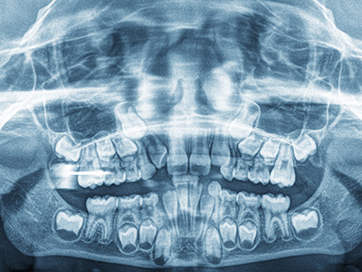
A cephalometric X‑ray is a specialized radiographic image that captures the entire head from the side, showing the relationship between the teeth, jaws, and skull. Unlike routine dental bitewings or panoramic radiographs, a cephalometric image provides a lateral profile that helps clinicians visualize skeletal patterns, tooth positions, and soft tissue contours in one plane. This single view is especially useful in orthodontic and growth‑related evaluations because it reveals how the skeletal and dental structures interact as a child develops.
This type of imaging is not intended to diagnose tooth decay; rather, it focuses on alignment, jaw relationships, and how the teeth sit within the facial skeleton. Orthodontists and pediatric dentists use cephalometric X‑rays to assess whether a child’s bite is balanced, whether there is a tendency toward underbite or overbite, and how facial proportions may change over time. The image serves as a baseline that can be compared with later images to track growth or treatment progress.
For parents, a cephalometric X‑ray may seem technical, but its purpose is straightforward: provide a reliable map of the facial skeleton so clinicians can make informed decisions about timing and type of treatment. When combined with a clinical exam and other imaging, it becomes a powerful tool for planning interventions that support healthy, functional development of the teeth and jaws.
Cephalometric X‑rays play a central role in treatment planning for children and adolescents because they reveal relationships that aren’t apparent from intraoral views alone. For example, two children might have similarly crowded teeth when viewed inside the mouth, yet their underlying jaw positions could be very different—one with a retrusive lower jaw and another with normal jaw alignment. Those differences change the approach to treatment, such as whether to focus on guiding jaw growth or on aligning teeth alone.
Timing is a major benefit of cephalometric analysis. Growth patterns differ widely between children, and a lateral skull image helps clinicians predict how much natural change is left. That prediction guides decisions about whether to intercept a developing problem early or to wait until growth slows. Early intervention can sometimes simplify later orthodontic work, while premature treatment may be unnecessary; cephalometrics helps find the right balance.
Beyond alignment, these images can also contribute to evaluating airway space and soft tissue relationships. In some cases, the X‑ray suggests whether a child’s breathing pattern or tongue posture could be influencing dental development. Although cephalometric imaging is only one component of a comprehensive assessment, it often uncovers clues that shape a more effective, individualized treatment plan.
The procedure for taking a cephalometric X‑ray is quick and noninvasive. The child stands or sits with their head stabilized while a machine rotates or aligns beside the head to capture the lateral profile. Preparation is minimal: the patient removes glasses, hair accessories, and any metal near the jaw to prevent image interference. The actual exposure typically lasts a fraction of a second, and modern digital systems capture the image instantly for review.
Safety is a key consideration, especially for children. cephalometric imaging uses a low dose of radiation—comparable to other dental radiographs—and practices follow the ALARA principle (As Low As Reasonably Achievable) to minimize exposure. Lead aprons and thyroid collars are commonly used when appropriate, and clinicians order cephalometric X‑rays only when they will influence diagnosis or treatment decisions.
Because the process is fast and nonthreatening, most children tolerate the exam easily. Technicians and dental staff take care to explain each step in kid‑friendly language and to position patients so the image is clear without repeated exposures. If a child feels anxious, the team can use comfort techniques to keep the experience calm and efficient.
Interpreting a cephalometric X‑ray involves identifying key landmarks on the skull and measuring angles and distances that describe jaw position, dental inclination, and facial proportions. Orthodontists use standardized analyses—such as SNA, SNB, and ANB angles—to quantify how the maxilla and mandible relate to the cranial base and to each other. Those measurements translate into practical treatment decisions, like whether to apply growth‑modification appliances or plan for fixed orthodontics.
Beyond numerical values, clinicians assess the soft tissue profile: lip position relative to the teeth, chin projection, and overall facial harmony. These observations are important because successful orthodontic care balances functional outcomes with facial aesthetics, particularly as a child matures. The cephalometric record provides an objective baseline against which future changes can be judged.
When treatment begins, follow‑up cephalometric images may be taken at key milestones to document progress and to fine‑tune the plan. This iterative approach helps ensure that interventions are responding to a child’s unique growth trajectory rather than a one‑size‑fits‑all protocol. Clear communication of findings helps families understand why a particular approach was chosen and what to expect during treatment.
Cephalometric X‑rays are ordered based on clinical need rather than routine habit. Common indications include initial orthodontic assessments, evaluation of jaw discrepancies, monitoring of growth for developing malocclusions, and pre‑surgical planning in complex cases. Because the image offers information that can change the course of care, clinicians weigh the benefit of the diagnostic insight against the need to limit radiation exposure.
The frequency of cephalometric imaging is individualized. In many cases, one image at the start of orthodontic planning and another near the end of active growth is sufficient. For children undergoing growth‑modification therapies or complex treatments, an intermediate image may be warranted to ensure the plan remains on track. Each decision is made with the child’s long‑term health and safety in mind.
Families should feel empowered to ask why an imaging study is recommended and how it will influence care. When ordered thoughtfully, a cephalometric X‑ray is a low‑risk procedure that yields high‑value information, helping clinicians deliver targeted, effective treatment while minimizing unnecessary interventions.
Cephalometric X‑rays are a fundamental diagnostic tool in pediatric orthodontics and play a vital role in shaping individualized, growth‑aware treatment plans. If you’d like to learn more about how this imaging is used for children and teens — or whether it may be appropriate for your child — please contact Myers Pediatric Dentistry & Orthodontics for more information.


A cephalometric x-ray is a lateral skull radiograph that captures the relationship among the teeth, jaws and cranial skeleton in a single profile view. Clinicians use this image to evaluate skeletal pattern, dental inclination and soft tissue contours that are not visible on intraoral films alone. The image provides an objective baseline that helps guide timing and type of orthodontic or growth-related interventions.
Because it shows the entire head in one plane, a cephalometric study is particularly helpful for understanding how a child’s facial proportions may change as they grow. It complements clinical examination and other imaging to produce a more complete diagnostic picture. When interpreted alongside growth data and dental models, the film supports individualized treatment planning that balances function and facial aesthetics.
Cephalometric x-rays are taken from the side of the head and emphasize skeletal relationships and facial proportions, while panoramic and bitewing radiographs focus on teeth, tooth roots and jawbone in different planes. Panoramic images show the entire dentition and jaw arches on a single curved plane, and bitewings are designed to detect interproximal decay and bone levels. Each radiograph has a different diagnostic role, and cephalometric images are chosen when skeletal and growth considerations are primary.
Because the cephalometric film captures craniofacial landmarks, it allows clinicians to perform standardized measurements such as SNA, SNB and ANB angles that quantify jaw relationships. Those measurements cannot be derived from intraoral films or panoramics. Using the right image for the clinical question improves diagnostic clarity and helps avoid unnecessary exposures.
Yes. Modern cephalometric imaging uses a low dose of radiation and practitioners follow the ALARA (As Low As Reasonably Achievable) principle to minimize exposure for pediatric patients. Lead aprons and thyroid collars are used as appropriate and digital systems often require lower exposure times than older film-based equipment. Clinicians recommend cephalometric imaging only when the expected diagnostic benefit outweighs the minimal radiation risk.
Technicians position the child carefully to get a clear image on the first attempt, which reduces the chance of repeat exposures. If parents have concerns about radiation, the dental team can explain the specific reasons for the study and how the information will influence care. Open communication helps families understand that safety measures are part of routine pediatric imaging protocols.
Preparation for a cephalometric x-ray is minimal and straightforward: the child removes glasses, hair accessories and any removable metal near the head and neck to prevent image artifacts. The staff will explain each step in age-appropriate language and will position the child with a forehead or chin rest to stabilize head posture. Because the exposure is very brief, there is no need for sedation or special fasting.
If your child is anxious, the dental team can use calming techniques or distraction to make the visit easier and avoid movement during the shot. Parents should let the staff know about any special needs or mobility concerns so positioning can be adjusted safely. Clear instructions and a gentle approach typically result in a fast, successful appointment with a single exposure.
Cephalometric x-rays are commonly recommended during initial orthodontic assessments to evaluate jaw relationships, growth patterns and the position of the teeth within the facial skeleton. They are especially useful when clinicians suspect skeletal discrepancies such as significant overbite, underbite or asymmetry that may require growth modification or more complex treatment. The study helps determine whether early intervention, monitoring or later comprehensive orthodontics is most appropriate.
In addition to initial planning, cephalometric imaging may be used when a clinician needs objective growth data to compare against normative values or to document treatment progress. The decision to order the image is individualized and based on whether the additional information will change management. This selective approach helps balance diagnostic value with prudent use of radiographs.
Interpreting a cephalometric x-ray involves identifying craniofacial landmarks and measuring angles and linear distances that describe skeletal and dental relationships. Common analyses include SNA and SNB to gauge maxillary and mandibular positions relative to the cranial base, and ANB to quantify jaw disharmony. Clinicians also evaluate dental inclinations, vertical proportions and soft tissue relationships such as lip position and chin projection.
Beyond numbers, the team assesses facial balance and functional factors that influence treatment choices, including airway space and tongue posture when visible on the lateral film. The radiograph becomes part of a larger diagnostic framework that includes clinical exam, dental casts and, when indicated, additional imaging. This integrated interpretation supports targeted, individualized care plans.
Frequency of cephalometric imaging is individualized according to clinical need rather than routine schedules. Many cases require only one image at the start of orthodontic planning and another near the end of active growth or treatment, while complex growth-modification cases may merit one or more intermediate films to verify progress. Each additional image is ordered only when it will meaningfully influence treatment decisions.
The timing also depends on the type of therapy being used and the child’s growth stage; for example, appliances intended to modify jaw growth often benefit from midpoint evaluation. Clinicians balance the need for information with radiation stewardship, and they will explain the rationale and timing for any follow-up imaging to families. Regular clinical exams remain central to monitoring between radiographs.
Cephalometric x-rays can provide useful information about upper airway space and the relationship of soft tissues in the oropharyngeal region, which may offer clues about breathing patterns or tongue posture. While the lateral film is not a definitive airway study like a sleep study or cone-beam CT, it can reveal anatomical tendencies that warrant further evaluation, such as reduced posterior airway space or a retrusive jaw posture. These findings may prompt referral or additional testing when breathing problems are suspected.
Because airway assessment often requires a multidisciplinary approach, cephalometric observations are combined with clinical history, examination and, when appropriate, collaboration with pediatricians or sleep specialists. Using the film as one piece of diagnostic information helps clinicians consider functional contributors to dental development and plan interventions that support both oral health and airway function.
A cephalometric x-ray provides important objective data about jaw relationships, dental inclination and facial proportions, which help clinicians determine whether orthodontic treatment or, in rare cases, orthognathic surgery should be considered. However, treatment recommendations are never based on a single image alone; clinicians integrate cephalometric findings with clinical examination, dental casts and growth assessment to form a comprehensive plan. The film clarifies whether the primary issue is dental alignment, skeletal discrepancy or a combination of both.
If skeletal concerns are significant enough to suggest surgical correction in the future, the cephalometric record becomes part of long-term planning and coordination with an oral and maxillofacial surgeon. For most children, early or phased orthodontic care guided by radiographic and clinical data can address problems without surgery. The dental team will discuss likely options, expected goals and monitoring steps so families understand the path forward.
At Myers Pediatric Dentistry & Orthodontics, cephalometric imaging is used selectively to inform growth-aware, individualized treatment plans for children and teens. We combine lateral radiographic analysis with a comprehensive clinical exam and discussion of developmental goals to decide whether early intervention, monitoring or full orthodontic treatment is most appropriate. The image helps our team quantify jaw relationships and document baseline measurements that guide appliance selection and timing.
When cephalometric studies are indicated, our clinicians explain the findings in clear terms so families understand how the information shapes care. Follow-up imaging is ordered only when it will change management, and all procedures follow pediatric safety protocols to minimize exposure. This measured, evidence-based approach supports predictable, child-centered outcomes and helps families feel confident in the plan.

Ready to schedule your child’s next dental visit or have questions about our services?
Contacting Myers Pediatric Dentistry & Orthodontics is simple! Our friendly team is here to help with scheduling appointments, explaining treatments, and answering any questions you may have. Whether you’d like to call, email, or use our easy online form, we’re ready to make your child’s dental experience positive and stress-free. Reach out today and give your little one a healthy, happy smile!

Back to top