New Patients
Existing Patients
New Patients
Existing Patients
New Patients
Existing Patients
New Patients
Existing Patients

Digital radiography replaces traditional film with sensitive electronic sensors and computer processing to capture images of teeth, gums, and supporting bone. Instead of waiting for film to develop, clinicians receive high-resolution images almost instantly, which improves the pace of care without sacrificing diagnostic detail. For families and patients, the experience is quicker and less disruptive; for clinicians, it provides a clearer window into oral health that supports timely, evidence-based decisions.
At its core, the technology converts X‑ray energy into digital data that can be adjusted, enlarged, and analyzed on-screen. These images retain the anatomical detail clinicians need to detect cavities, evaluate root and bone structure, and monitor growth and development in younger patients. Because the images are digital files rather than physical negatives, they also integrate seamlessly with electronic health records and other diagnostic tools.
Digital radiography is especially well suited for pediatric dentistry because speed and clarity reduce the need for repeat exposures and help children stay calm during appointments. When combined with a gentle approach and age-appropriate communication, digital imaging supports a positive visit while still giving clinicians the information necessary to protect long-term dental health.
One of the most important benefits of digital radiography is its efficiency at using lower doses of radiation compared with older film techniques. Advances in sensor sensitivity and image-processing algorithms mean clinicians can obtain diagnostic-quality images with less exposure. This reduction is particularly meaningful for children, who are more sensitive to ionizing radiation and may require repeat imaging over the years as their mouths develop.
Digital systems also make it easier to follow modern guidelines for when X‑rays are clinically necessary. Rather than applying a one-size-fits-all approach, pediatric dentists assess each child's risk and tailor imaging intervals to individual needs. The result is a balanced strategy that prioritizes safety while still catching issues early enough to simplify treatment and preserve dental health.
Beyond the technical reduction in radiation dose, digital workflows reduce the likelihood of retakes caused by processing errors, underexposure, or unclear images. That reliability means fewer repeats, less cumulative exposure, and greater reassurance for parents who want strong safety standards for their children’s care.
Digital images offer fine-grain detail and the ability to enhance contrast, crop, and magnify specific areas without degrading image quality. These capabilities help clinicians detect early-stage cavities between teeth, evaluate the integrity of restorations, and assess developing tooth roots and bone levels. Image-enhancement tools can highlight subtle changes that might be missed on film, supporting more precise clinical judgments.
Because digital files can be viewed immediately on a monitor, clinicians and parents can review findings together in real time. Visual explanations help families understand a proposed care plan and see the reason behind recommended preventive steps or treatments. This visual communication often improves cooperation and follow-through, especially with children who respond well to simple, visual demonstrations.
For complex cases, digital radiography integrates with other diagnostic technologies — for example, intraoral cameras and three-dimensional imaging — allowing clinicians to form a more complete clinical picture. That interoperability contributes to higher-quality care and better-coordinated treatment planning across different modalities.
Digital radiography streamlines clinical workflow from capture to storage. Images are saved directly into a patient’s electronic record, eliminating darkrooms, chemical processing, and the physical storage space required for film. Staff can retrieve prior images instantly, compare them side-by-side, and track changes over time, which makes recall visits and monitoring much more effective.
Because files are easily shared in digital format, consultations with specialists or transfers between offices happen quickly and securely. When a pediatric dentist coordinates with an orthodontist, oral surgeon, or primary physician, digital images can be transmitted without delay, speeding up referrals and ensuring everyone involved in a child’s care sees the same diagnostic information.
From an operational standpoint, digital imaging reduces administrative friction and improves documentation quality. Clear, time‑stamped images in the electronic record support clinical continuity and help providers make consistent, well-documented decisions as a child’s dental needs evolve.
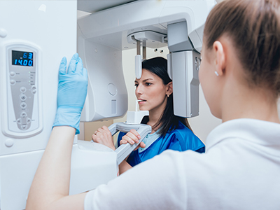
Digital radiography appointments are typically brief and designed with comfort in mind. When an X‑ray is needed, a small sensor is placed intraorally or positioned externally depending on the image required. The sensor is sized and positioned with extra attention for pediatric patients, and staff use gentle techniques to reduce movement and help children relax. The actual exposure lasts only a fraction of a second, and because images appear immediately on the screen, there’s no waiting for development.
Parents are often invited to view images with the dental team so they can see what the clinician sees and participate in treatment discussions. The ability to explain findings visually makes it easier to answer questions and set expectations for preventive measures or next steps. Clinicians will always balance the need for imaging with the principle of using the least amount of radiation necessary to obtain diagnostic information.
After the visit, digital images remain part of the child's record and are available for future comparisons. This continuity helps detect trends and guide long-term care — from routine preventive visits to more involved restorative or orthodontic planning — while maintaining a focus on safety and minimally invasive treatment whenever possible.
Digital radiography brings faster imaging, enhanced diagnostic clarity, and improved safety to pediatric dental care. By reducing radiation exposure, enabling immediate image review, and simplifying recordkeeping, it supports better outcomes for growing smiles. The technology also strengthens teamwork between providers and families through clear visual communication and efficient information sharing.
At Myers Pediatric Dentistry & Orthodontics, our team uses digital imaging as one of several modern tools to provide thoughtful, child-centered care. If you have questions about how digital X‑rays are used during an appointment or want to learn how imaging fits into your child’s care plan, please contact us for more information.


Digital radiography uses electronic sensors and computer processing to capture X-ray images instead of photographic film. The sensors convert X‑ray energy into digital data that can be viewed, enhanced, and stored instantly on a computer. This immediacy speeds diagnosis and removes the need for chemical processing and physical film storage.
Because images are digital files, clinicians can enlarge, adjust contrast, and compare shots side by side without degrading detail, which improves diagnostic clarity. Digital files also integrate with electronic health records and other diagnostic tools, making it easier to track changes over time. For pediatric care, the faster workflow helps keep visits calm and efficient for children and families.
Digital radiography typically requires lower radiation doses than older film techniques because modern sensors are much more sensitive and image-processing software can enhance detail. Dentists follow the ALARA principle — keeping exposure As Low As Reasonably Achievable — and tailor imaging to each child’s needs to limit unnecessary X‑rays. This individualized approach reduces cumulative dose while preserving diagnostic value.
Because children can be more sensitive to ionizing radiation and may need periodic imaging as they grow, the combination of sensitive sensors and careful clinical judgment is especially important in pediatric dentistry. Staff also take practical steps to minimize exposure, such as using fast sensors, proper shielding, and positioning techniques to avoid retakes. The result is a safer imaging process that supports long-term oral health monitoring.
Digital images provide high-resolution detail and tools for enhancement, such as contrast adjustment and magnification, which help clinicians spot early cavities and subtle changes in root or bone structure. These capabilities allow for earlier intervention when problems are easier to treat and help clinicians plan restorations or orthodontic care with greater precision. Viewing images immediately also supports clearer, evidence-based decisions during the visit.
When needed, digital radiography integrates with other imaging technologies like intraoral cameras and three-dimensional scans to form a comprehensive diagnostic picture. That interoperability improves coordination between pediatric dentists, orthodontists, and other specialists, enabling more accurate referrals and better-managed treatment sequences. Visual images also help families understand recommended care and follow-through on preventive or restorative plans.
There is no single timetable that fits every child; recommended imaging intervals depend on a child’s age, dental development, and risk factors such as a history of cavities or orthodontic needs. Pediatric dentists assess each child individually and follow professional guidelines to determine whether bitewings, periapical, panoramic, or other images are clinically necessary. The goal is to balance early detection with minimizing exposure.
For very young or low-risk children, X‑rays may be less frequent and focused on developmental milestones, while children with active decay or orthodontic planning may need periodic imaging to monitor progression. Clinicians review prior images and clinical findings to decide when a new image will add meaningful information. Parents are encouraged to ask about the rationale for any recommended films so they understand how imaging fits into their child’s care plan.
Appointments are usually brief and designed with comfort in mind: a small sensor is placed in the mouth or the machine is positioned externally, and the exposure itself lasts only a fraction of a second. Staff use pediatric-sized sensors, gentle positioning techniques, and clear, age-appropriate instructions to reduce movement and help children stay relaxed. Because images appear on-screen immediately, there is no film development wait time.
Parents are often invited to view images with the dental team so they can see findings and discuss next steps in real time. Clinicians explain what each image shows and how it relates to preventive care or any recommended treatment, helping families make informed decisions. After the visit, digital images become part of the child’s record and are available for future comparison.
Immediate on-screen review lets clinicians confirm image quality right away, so a retake can be avoided or performed immediately if necessary rather than after film is developed. Modern sensors and processing algorithms also reduce errors caused by underexposure or processing faults that were common with film. Accurate positioning aids and pediatric-specific techniques further lower the chance of blurred or unusable images.
Reducing retakes lowers a child’s cumulative exposure and shortens appointment time, which improves the overall patient experience. Staff training in pediatric imaging protocols and routine equipment calibration help maintain consistent image quality. Together these factors create a more reliable workflow and greater reassurance for families about safety standards.
Yes. Digital images are available for parents to view during the appointment so the dental team can explain findings visually and discuss preventive steps or treatments. Seeing the images together helps families understand the clinical reasoning behind recommendations and supports informed decision-making. Visual explanations are especially helpful when discussing cavity locations, growth patterns, or the need for monitoring.
Images are stored in the child’s electronic record for future comparison and can be shared with other providers if parents request a specialist consultation. When images are shared, clinicians follow secure transfer practices to ensure records are transmitted appropriately. Parents who want copies or a detailed explanation should speak with the front office or the dental team after the appointment.
Digital radiographs are saved directly into a patient’s electronic chart with clear time stamps, which streamlines recordkeeping and makes prior images easy to retrieve for comparison. Electronic storage eliminates physical film archives and supports consistent documentation of changes over time. Staff follow privacy and security procedures to protect health information stored in these systems.
When images are shared with specialists or other offices, they are transmitted using secure methods and with appropriate permissions to maintain confidentiality. This secure sharing improves care coordination for treatments like orthodontics or surgical referrals without the delays associated with physical film. Clear documentation of images in the chart also supports continuity of care as a child’s dental needs evolve.
Digital radiography includes a variety of image types: intraoral bitewings and periapicals for detailed views of individual teeth and surrounding bone, panoramic images for a broad view of the jaws and developing teeth, and three-dimensional cone-beam scans for complex diagnostic or surgical planning. Bitewings are commonly used to check for cavities between teeth, while periapicals evaluate tooth roots and surrounding bone. Panoramic and CBCT images are used selectively when a wider or 3D perspective is clinically necessary.
In pediatric dentistry, clinicians choose the least intrusive image that will answer the clinical question, reserving advanced 3D scans for cases that require detailed spatial information. That selective use aligns with safety principles while ensuring clinicians have the imaging needed for accurate diagnosis and treatment planning. Parents can ask the dental team which type of image is recommended and why it is appropriate for their child.
Myers Pediatric Dentistry & Orthodontics uses digital radiography because it delivers faster results, high diagnostic clarity, and lower radiation exposure compared with older film methods. The technology supports child-centered care by shortening appointment time, reducing retakes, and enabling visual explanations that help families understand treatment recommendations. These benefits align with the practice’s emphasis on gentle, evidence-based pediatric dentistry.
Digital imaging also improves recordkeeping and collaboration with specialists when coordinated care is needed at the Middleburg or Jacksonville locations. Storing images in electronic charts helps clinicians track growth and treatment outcomes over time, supporting thoughtful, long-term oral health planning for growing smiles. Families with questions about how imaging fits into a specific care plan are encouraged to discuss their concerns with the dental team during the appointment.

Ready to schedule your child’s next dental visit or have questions about our services?
Contacting Myers Pediatric Dentistry & Orthodontics is simple! Our friendly team is here to help with scheduling appointments, explaining treatments, and answering any questions you may have. Whether you’d like to call, email, or use our easy online form, we’re ready to make your child’s dental experience positive and stress-free. Reach out today and give your little one a healthy, happy smile!

Back to top