New Patients
Existing Patients
New Patients
Existing Patients
New Patients
Existing Patients
New Patients
Existing Patients

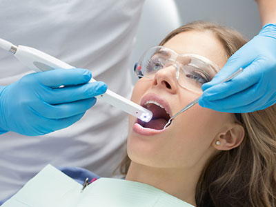
An intraoral camera is a compact, pen-sized imaging device that captures high-resolution, full-color pictures from inside the mouth. For pediatric patients, this means dentists can show a detailed view of teeth, gums, and other soft tissues on a monitor in real time. The level of detail makes it easier to spot early signs of decay, cracks, enamel wear, or areas where brushing may be missed—issues that may be hard to detect with the naked eye alone.
Unlike abstract descriptions or drawings, live images from an intraoral camera let parents and patients see exactly what the dentist sees. This visual clarity builds understanding and helps clinicians explain the nature and location of a problem without relying solely on technical language. For kids, seeing a friendly, illuminated image of their own teeth can also reduce anxiety by turning an unseen process into something familiar and explainable.
The camera’s small size and gentle lighting are designed with comfort in mind. It slips easily around small mouths and between teeth, producing crisp images quickly so examinations remain short and minimally invasive. Because the images are digital, they can be immediately reviewed, saved to the patient record, and referenced during follow-up visits to track changes over time.
High-quality intraoral photographs improve diagnostic accuracy by providing views from angles that are difficult to achieve during a routine clinical exam. Dentists can enlarge images to inspect suspicious spots, compare surfaces side by side, and document findings with precision. This additional visual information complements traditional examinations and radiographs, helping clinicians make more informed decisions about when and how to intervene.
For treatment planning, these images serve as an objective record that clarifies the exact location and extent of an issue. Whether deciding on a protective sealant, a small restoration, or more comprehensive care, the intraoral camera helps the dental team prioritize interventions that preserve tooth structure and support long-term oral health. Clear images also reduce guesswork and provide a consistent reference point when multiple providers collaborate on a case.
Because the camera captures still images and video, it can be used during procedures to confirm outcomes in real time. This capability is especially helpful in pediatric dentistry, where minimally invasive approaches and conservative restorations are preferred. Being able to show parents what was treated—and why—builds transparency and supports shared decision-making between clinicians and caregivers.
A typical intraoral camera exam is straightforward and child-friendly. The dentist or hygienist will explain the tool in simple terms, often letting a child hold the device briefly before it is used so they feel more comfortable. During the exam, the camera is passed gently over the teeth and gums; its small size allows for quick captures without causing discomfort or requiring wide mouth opening for long periods.
Images appear almost instantly on a monitor, where the clinician can point out areas of interest and describe what the picture shows. Parents are encouraged to watch and ask questions; seeing the images together makes explanations more concrete and helps caregivers understand how daily habits—like brushing and flossing—affect oral health. The process is designed to be collaborative and educational rather than intimidating.
Following the exam, selected images are saved into the patient’s record for future comparison. If additional evaluation is necessary, the practitioner will outline next steps clearly, using the images as a reference. This helps families know exactly why a recommendation is being made and what outcomes the team expects with timely care.
Intraoral imaging benefits everyone involved in a child’s dental care. For parents, the visual evidence supports clearer communication about oral hygiene needs and upcoming treatments, making it easier to follow through with home care instructions. For specialists and laboratories, shared images provide precise visual documentation that streamlines referrals and collaborative treatment planning.
These saved images also strengthen the continuity of care. When a child moves between providers or returns after a period of growth, historical photos enable clinicians to see how conditions have evolved. That historical perspective is especially important in pediatric dentistry, where teeth and mouths change rapidly as children grow and permanent teeth erupt.
Digital images are also useful for monitoring preventive measures over time. Clinicians can compare images from different visits to confirm that a sealant, restoration, or hygiene routine is performing as intended. This ongoing visual record supports proactive care and helps avoid unnecessary interventions by catching changes early.
Modern intraoral cameras combine bright, shadow-free LED illumination with high-resolution sensors to produce clear, accurate images. Many systems include software tools for annotating, magnifying, and archiving photos, enabling clinicians to highlight areas of concern and track changes across appointments. These features are integrated into secure dental record systems so images remain part of the patient’s official chart.
From a safety standpoint, intraoral cameras are non-invasive and rely on visible light only. Devices used in clinical settings are designed for repeated patient contact and follow strict infection-control protocols; disposable sleeves or sterilizable tips are used when appropriate, and instruments are cleaned according to accepted guidelines between patients. The result is a low-risk diagnostic tool that adds significant value to routine care.
Clinicians who use intraoral cameras do so as part of a broader diagnostic toolkit that includes clinical examination and radiography when indicated. The images are intended to enhance—not replace—professional judgment. When combined with training, experience, and other diagnostic methods, intraoral imaging helps dental teams deliver care that is more precise, transparent, and patient-centered.
Wrap-up: Intraoral cameras bring clear, objective imaging into pediatric dental care, improving diagnosis, communication, and long-term monitoring. At Myers Pediatric Dentistry & Orthodontics we incorporate this technology to help patients and parents see what the dentist sees and to support thoughtful, conservative treatment choices. Contact us to learn more about how intraoral imaging may be used during your child’s visit and to discuss any questions you have about the process.


An intraoral camera is a small, pen-sized imaging device that captures high-resolution, full-color photos and video from inside the mouth. It uses a bright, shadow-free LED light and a high-resolution sensor to produce clear images of teeth, gums and soft tissues. Images are transmitted digitally to a monitor so clinicians, parents and patients can view them in real time.
The camera head is compact and designed to navigate small mouths, allowing quick captures from angles that may be hard to see by eye. Captured images can be enlarged, annotated and saved to the patient record for comparison over time. This immediate visual feedback helps clinicians explain findings clearly and document conditions objectively.
Intraoral images reveal early signs of decay, cracks, enamel wear and areas where plaque accumulates that may be missed during a routine visual exam. High magnification and alternative viewing angles let clinicians inspect suspicious surfaces with greater precision. This level of detail improves diagnostic accuracy and supports timely, conservative interventions when needed.
Images complement clinical examination and radiographs by providing surface-level detail and color information that X-rays do not show. Dentists can compare photos side by side, track progression and document subtle changes between visits. For pediatric patients, visual records are especially helpful because mouths change quickly as primary teeth fall out and permanent teeth erupt.
Yes, intraoral cameras are noninvasive devices that use visible LED light and digital sensors rather than radiation. They pose no known health risks when used as intended and are considered safe for patients of all ages. The small, rounded camera heads and soft lighting are designed to minimize discomfort during use.
Clinical use follows infection-control protocols, including disposable sleeves or sterilizable tips and routine cleaning between patients. These measures prevent cross-contamination and maintain a safe environment for children. When combined with standard sterilization practices, intraoral imaging remains a low-risk diagnostic tool.
The clinician will explain the device in simple terms and often show the monitor so the child and parent know what to expect. The camera is then passed gently over the teeth and gums while images appear almost instantly on the screen. Captures are quick, typically taking only a few seconds per view, which helps keep exams short and child-friendly.
Parents are encouraged to watch the live images and ask questions so explanations are concrete and collaborative. Selected photos are saved to the chart for future comparison and to support treatment planning when necessary. In our Middleburg and Jacksonville offices the process is integrated into routine visits and tailored to each child's comfort level.
No, intraoral images and dental X-rays serve different diagnostic purposes and are complementary tools. Cameras excel at showing surface detail, color and soft-tissue conditions, while X-rays reveal structures below the gumline and between teeth, such as interproximal decay and bone levels. A comprehensive assessment typically uses both methods when clinically indicated.
Clinicians use intraoral photos to document visible findings and guide discussions, then rely on radiography for information that cannot be seen externally. Together, the two approaches enable a fuller understanding of a child's oral health and support informed treatment decisions. Your dental team will recommend the appropriate combination of exams based on your child's individual needs.
High-quality images provide objective documentation of the exact location and extent of a problem, making it easier to prioritize conservative care. Clinicians can annotate, magnify and compare photos to explain why a sealant, restoration or further evaluation is recommended. Visual evidence reduces ambiguity and supports shared decision-making between providers and families.
At Myers Pediatric Dentistry & Orthodontics we use intraoral imaging to show parents clear images of treated areas and to demonstrate proper home-care techniques. This transparency helps caregivers understand the rationale for each recommendation and what to watch for at home. Saved images also provide a reference during follow-up visits to confirm that interventions are performing as intended.
Yes, clinicians often encourage children to look at the monitor so they understand what is being examined and feel more involved. If appropriate, a team member may allow the child to hold the device briefly to reduce anxiety and build familiarity. This hands-on introduction is always supervised and followed by a gentle, professional exam.
Allowing a child to participate can transform an unfamiliar instrument into a predictable part of the visit, which helps reduce fear. The clinical team remains focused on safety and speed, keeping captures brief to maintain comfort. Parents can ask the staff how they tailor the experience for younger or more anxious children.
Digital images are integrated into the secure electronic patient record and labeled with the exam date and relevant notes for easy retrieval. Access is restricted to authorized clinical staff and handled according to privacy and security policies. These practices help maintain continuity of care while protecting patient information.
At Myers Pediatric Dentistry & Orthodontics images are stored within our practice management system so clinicians can compare visits over time and share visuals with specialists when needed. When images are shared, clinicians follow secure transfer protocols and obtain parental consent as required. Parents can ask the office about specific privacy practices or how long images are retained.
Yes, cameras can capture still photos or video during and after procedures to document the treatment outcome in real time. This is useful for verifying the fit of a restoration, the placement of a sealant, or the completion of a conservative procedure. Real-time imaging supports clinical judgment and provides an objective record of the result.
Using intraoral photos during treatment also helps clinicians explain the procedure to parents and demonstrate what was accomplished. Saved images become part of the chart and can be referenced in follow-up visits to confirm healing and durability. This documentation enhances transparency and helps the dental team monitor clinical success over time.
Key features to consider include high-resolution sensors, bright LED illumination and software that allows enlargement and annotation of images. Good integration with the practice's patient record system ensures photos are saved and accessible for future comparison. Parents should also inquire whether the practice follows strict infection-control protocols for camera use.
A practice that explains images clearly, encourages questions and uses photos as part of education demonstrates a patient-centered approach to care. When combined with experienced clinicians and appropriate diagnostic tools, intraoral imaging becomes a practical aid for monitoring growth and catching issues early. Asking about how images are used during visits can help families understand the value of the technology.

Ready to schedule your child’s next dental visit or have questions about our services?
Contacting Myers Pediatric Dentistry & Orthodontics is simple! Our friendly team is here to help with scheduling appointments, explaining treatments, and answering any questions you may have. Whether you’d like to call, email, or use our easy online form, we’re ready to make your child’s dental experience positive and stress-free. Reach out today and give your little one a healthy, happy smile!

Back to top