New Patients
Existing Patients
New Patients
Existing Patients
New Patients
Existing Patients
New Patients
Existing Patients

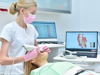
The iTero® Scanner captures a detailed three-dimensional map of the teeth and surrounding soft tissues without using traditional impression trays and putty. Instead of waiting for impression material to set, the device records a continuous digital scan that is immediately viewable on a monitor. That digital model serves as the foundation for diagnostic evaluations, orthodontic planning, restorative designs, and many other clinical workflows.
Because the scan creates a high-resolution virtual impression, clinicians can examine angles and details that are hard to see with conventional impressions. The data can be rotated, zoomed, and compared to previous scans to monitor changes over time. This visual clarity helps dental teams identify issues earlier and make more informed recommendations for preventive or corrective care.
Clinicians also benefit from standardized digital records. A single scan can be stored, duplicated, and shared with labs or specialists without degradation, which reduces errors associated with physical impressions. For practices committed to modern workflows, the iTero® Scanner becomes a central tool for accurate, repeatable treatment planning.
Because the scanning data integrates with many digital systems—aligner manufacturers, CAD/CAM labs, and practice management software—its versatility speeds coordination across the care team. That interoperability makes the iTero® system especially valuable for pediatric and orthodontic practices focused on efficient, predictable outcomes.
One of the clearest benefits patients notice is comfort. Traditional impressions can be uncomfortable for children and adults alike—tray gagging, sticky material, and a lingering taste are common complaints. A digital scan avoids those issues by using a small, hand-held wand that glides along the dental arches while the patient breathes and swallows normally.
For families, the absence of messy impression materials translates into a more positive appointment experience. Young patients who might otherwise feel anxious about dental procedures often respond better to a noninvasive, quick scanning process. The clinician can pause, review, and repeat brief segments if needed without subjecting the patient to prolonged discomfort.
Beyond comfort, digital scanning generally reduces the time spent in the chair. Quick capture and instant review eliminate the wait associated with setting impressions and shipping physical models. For busy families and children with limited patience or special needs, shorter, calmer visits make a meaningful difference in overall care adherence.
That ease of experience helps the dental team focus on communication and education. With real-time images on-screen, clinicians can show parents and patients precise views of areas of concern and explain recommended treatments in plain language, increasing understanding and trust.
Accuracy is a cornerstone of effective dental treatment, and digital scans deliver consistent, millimeter-level detail that supports precise restorative and orthodontic work. The iTero® Scanner captures occlusal relationships and soft-tissue contours that labs and aligner manufacturers use to fabricate appliances that fit predictably the first time.
For orthodontic cases, digital models enable virtual simulations that illustrate expected tooth movements. Clinicians can present a step-by-step plan and adjust parameters on-screen before any appliance is made. This proactive planning reduces the likelihood of remakes and adjustments later in treatment.
Restorative dentistry benefits from the same precision: crowns, bridges, and onlays designed from accurate digital impressions tend to seat better and require fewer intraoral adjustments. The cumulative effect is fewer return visits for refinements and a smoother continuum from diagnosis to final restoration.
Speed matters not only for patient convenience but for clinical efficiency. The ability to capture and transmit a digital impression immediately accelerates lab workflows and shortens fabrication times for restorations and aligners. Faster turnaround supports timely treatment starts, which is particularly important when working around school schedules and family commitments.
The iTero® Scanner uses non‑ionizing light-based imaging rather than X-rays to capture surface detail, so it does not expose patients to radiation during the scanning process. Because the device focuses on external dental surfaces and soft tissue contours, it complements radiographic imaging rather than replacing it when internal anatomy must be assessed.
This safety profile is especially appealing for families who prefer to limit unnecessary radiation exposure in routine procedures. Scanning is quick and repeatable, which allows clinicians to update digital records over time without concern about cumulative dose from the scanning process itself.
In practice, that means dental teams can monitor development and treatment progress frequently and comfortably. Whether tracking tooth eruption patterns in a growing child or evaluating fit and progress during orthodontic care, non‑radiographic scans provide a safe way to gather high-quality visual data at multiple appointments.
From a workflow perspective, the scanner’s ergonomics and ease of use reduce operator fatigue and minimize the need for retakes. Its handheld wand is designed to navigate pediatric mouths efficiently, allowing technicians and dentists to capture full-arch data even in patients who have limited tolerance for lengthy procedures.
Digital impressions create a shared visual language for the clinical team, the patient, and external partners such as labs or orthodontic manufacturers. Because the iTero® output is a manipulable 3D model, everyone involved sees the same anatomy and can collaborate more effectively on design and treatment sequencing.
This clarity matters when coordinating restorations or clear aligner therapy. Scans can be sent electronically, feedback can be integrated quickly, and final appliances are often more accurate because they’re based on a precise digital starting point. The result is less back-and-forth and fewer surprises at delivery.
Additionally, real-time visuals support clearer conversations with parents and older children. Clinicians can mark areas of interest on the screen, compare scans from different visits, and walk families through the expected steps of care. That transparent communication helps set realistic expectations and improves shared decision-making.
Practically speaking, implementing digital scanning elevates the overall standard of care by combining better diagnostic detail, improved patient comfort, and streamlined collaboration. Our team at Myers Pediatric Dentistry & Orthodontics incorporates this technology to support reliable, child‑centered care and to make the path from evaluation to treatment as smooth as possible.
In summary, the iTero® Scanner replaces messy impressions with fast, accurate, and patient-friendly digital scans that improve diagnostics, communication, and clinical efficiency. If you’d like to learn more about how digital scanning fits into routine pediatric or orthodontic care, please contact us for more information.


The iTero® scanner is a handheld intraoral imaging device that captures a detailed three-dimensional map of the teeth and surrounding soft tissues. It records continuous digital impressions using light-based imaging rather than traditional trays and putty, producing a high-resolution virtual model in real time. Clinicians can view the scan immediately on a monitor and capture any missed segments during the appointment.
The digital model created by the scan serves as the foundation for diagnostics, orthodontic planning, and restorative designs. Because files are stored digitally, they can be duplicated, compared to prior scans, and shared with labs or specialists without loss of fidelity. This capability supports repeatable treatment planning and reduces errors associated with physical impressions.
Digital scanning eliminates the need for impression trays and setting materials, which removes common issues such as tray discomfort, material distortion, and delayed feedback. Scans deliver consistent, millimeter-level detail for occlusal relationships and soft-tissue contours that are difficult to reproduce reliably with physical impressions. The immediacy of a digital model also allows clinicians to verify capture quality and repeat brief segments if necessary.
From a lab and workflow perspective, digital files transmit instantly to manufacturers and labs, reducing shipping time and the risk of physical damage. That streamlined transfer often decreases the need for remakes and intraoral adjustments at delivery. While radiographs remain necessary for internal anatomy, digital surface scans complement imaging by providing highly accurate external detail.
The iTero® scanner uses a small, handheld wand that glides along the dental arches while the patient breathes and swallows normally, making the process noninvasive and generally well tolerated by children. Because there is no messy impression material, patients avoid common complaints such as gagging or lingering tastes. Clinicians can pause and repeat brief segments if a child needs a short break, which helps accommodate varying tolerance levels.
The scanning technology is light-based and non‑ionizing, so it does not expose patients to radiation during the capture process. That safety profile allows clinicians to update digital records frequently without concern about cumulative dose from the scan itself. Additionally, the wand is ergonomically designed to navigate smaller mouths, which can shorten capture time and reduce operator fatigue.
Digital scans enable virtual simulations that illustrate expected tooth movements and treatment outcomes, giving clinicians a precise platform to plan and adjust treatment parameters before appliances are fabricated. With manipulable 3D models, providers can present step-by-step plans, show projected results, and optimize aligner staging or bracket positioning on-screen. This proactive planning reduces the likelihood of remakes and unnecessary adjustments during therapy.
During active orthodontic treatment, periodic scans allow teams to monitor progress objectively by comparing current models to the original plan. These comparisons help identify deviations early and guide midcourse corrections when needed. The result is a more predictable pathway from diagnosis to the desired alignment outcome.
Yes. The high-resolution digital impressions that the iTero® scanner produces are used for restorative workflows such as crowns, bridges, onlays, and inlays. Accurate capture of occlusal relationships and preparation margins helps dental technicians design restorations that fit more precisely at seating. Better initial fit typically reduces chairside adjustment time during delivery.
Because digital files transmit directly to CAD/CAM labs, the turnaround for restorations can be faster and more predictable than traditional cast-based workflows. The scanner also supports documentation of preexisting anatomy and posttreatment outcomes, which is useful for complex restorative cases. Overall, digital impressions contribute to a smoother continuum from preparation to final restoration.
A full-arch digital scan is commonly captured in just a few minutes, while shorter targeted scans require even less time depending on the area of interest. During the appointment a clinician or trained technician will move the wand around the dental arches while keeping the patient comfortable and able to breathe normally. The on-screen model assembles in real time so the team can confirm capture quality and briefly retake any areas that need refinement.
Patients should expect a calm, noninvasive process without impression trays or putty. Children with limited tolerance can be scanned in segments with short pauses between passes to maintain comfort. Because the clinician can review the data immediately, the visit often avoids delays associated with setting materials and shipping physical models.
The frequency of digital scans depends on the clinical purpose: initial baseline scans are helpful before orthodontic evaluation or restorative treatment, while monitoring scans are scheduled based on growth, treatment stage, or specific concerns. For growth and development tracking, clinicians commonly plan follow-up scans at clinically relevant intervals to document eruption patterns and arch changes. During active orthodontic therapy, scans may be taken at key milestones to verify progress and adjust treatment as needed.
Because the scanner uses non‑ionizing imaging, scans can be repeated safely over time to build a chronological record. The exact interval should be determined by the dental team in consultation with the family, balancing the value of updated visual data with the child’s comfort and appointment schedule. Documentation from periodic scans helps guide timely interventions and informed decision-making.
The iTero® system produces standardized digital files that are compatible with many CAD/CAM labs and aligner manufacturers, enabling direct electronic transmission of precise anatomy. This interoperability removes the need to ship physical models and reduces the risk of distortion or damage during transport. Labs and manufacturers use the high-resolution data to design appliances and restorations that fit more predictably on delivery.
For clear aligner therapy, digital scans can be used to generate virtual treatment simulations and to submit cases for manufacturing without intermediary physical impressions. Feedback from labs or manufacturers is often exchanged electronically, which shortens turnaround times and streamlines coordination across the care team. The net effect is a more efficient workflow from capture to final appliance or restoration.
Real-time 3D visuals create a shared reference that helps clinicians explain findings and recommended treatments in clear, concrete terms. On-screen models can be rotated, zoomed, and annotated so parents and older children see the same anatomy the team is reviewing, which improves understanding of issues and proposed next steps. Visual comparisons between visits also help illustrate growth or treatment progress in a way that is easier to interpret than verbal descriptions alone.
Using the scan as an educational tool supports informed consent and shared decision-making by making treatment options and expected outcomes more transparent. Clinicians can mark areas of interest, demonstrate how appliances will fit, and answer questions using the live model. This approach helps families feel more engaged and informed about their child’s care plan.
Myers Pediatric Dentistry & Orthodontics incorporates iTero® scans into the patient record to support diagnosis, treatment planning, and coordination with specialists and labs. Digital files are stored as part of the clinical chart and can be referenced to monitor development, confirm restorative margins, or transmit precise anatomy for appliance fabrication. Electronic transfer of scan data improves collaboration and reduces the turnaround time for ordered appliances and restorations.
The practice uses repeat scans to document treatment progress and to verify outcomes at critical stages, enabling timely adjustments when indicated. Scan records are managed in accordance with applicable privacy and record-keeping standards and are shared with external partners only with appropriate consent. This integration of digital scanning helps the team deliver consistent, child-centered care from initial evaluation through final treatment.

Ready to schedule your child’s next dental visit or have questions about our services?
Contacting Myers Pediatric Dentistry & Orthodontics is simple! Our friendly team is here to help with scheduling appointments, explaining treatments, and answering any questions you may have. Whether you’d like to call, email, or use our easy online form, we’re ready to make your child’s dental experience positive and stress-free. Reach out today and give your little one a healthy, happy smile!

Back to top