New Patients
Existing Patients
New Patients
Existing Patients
New Patients
Existing Patients
New Patients
Existing Patients

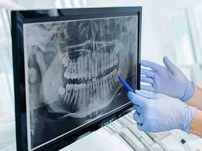
A panoramic X-ray is a single, wide-view radiograph that captures the entire mouth in one image — upper and lower teeth, both jaws, the temporomandibular joints (TMJ), and adjacent structures such as the maxillary sinuses. Rather than focusing on one tooth or a small area, it provides a broad overview of how the dental and jaw anatomy relate to each other. That big-picture perspective helps clinicians identify patterns and conditions that might be missed on isolated intraoral films.
Because the image displays the spatial relationships across the jaws, panoramic radiographs are especially useful for screening developmental issues and planning coordinated care. For children and adolescents, this means monitoring tooth eruption, identifying missing or extra teeth, and recognizing how skeletal growth will affect future alignment. In many cases a panoramic X-ray is the first step in a diagnostic process that guides safe, efficient care.
Beyond initial screening, panoramic images are valuable as a baseline record. They document the patient’s oral health at a given point in time, making it easier to track changes over months or years. That record is particularly helpful when coordinating care across specialties, such as when orthodontic evaluation or referral is part of a child’s treatment plan.
The panoramic X-ray process is designed to be simple and comfortable for patients of all ages. During the scan, the patient stands or sits and gently bites on a small guide to stabilize the head. A rotating arm moves around the patient’s head while an X-ray source and detector capture the full curved image of the jaws. The entire exposure takes less than a minute, and most of that time is positioning — actual radiation exposure lasts only a few seconds.
For children, the team will use age-appropriate instructions and positioning aids to ensure a clear image with minimal movement. The staff routinely works with younger patients to reduce anxiety: explaining each step, demonstrating equipment, and providing reassuring cues throughout the brief procedure. No sedation or invasive preparation is required for a standard panoramic exam.
Digital panoramic units produce images instantly, allowing the clinical team to review results with families at the same visit. That rapid feedback helps clinicians explain findings clearly and outline next steps without delay, which improves communication and supports timely decision-making for preventive or restorative care.
In pediatric dentistry and orthodontics, panoramic X-rays play a central role in assessing development and planning treatment. They show unerupted permanent teeth, retained baby teeth, supernumerary (extra) teeth, and congenitally missing teeth. By revealing the position and angulation of developing teeth, panoramics help anticipate crowding, impaction, or spacing issues before they become more complex.
Orthodontists frequently rely on panoramic images to evaluate the relationship between the dental arches and to decide whether early intervention is advisable. The image helps determine if space management, growth modification, or referral for appliances is needed. It also assists in tracking jaw symmetry and the timing of key developmental milestones that influence orthodontic timing.
Panoramic radiographs are also useful for clinical situations such as assessing trauma, evaluating signs of infection that may involve multiple teeth or the jaw, and screening for cysts or other abnormalities. While they don’t replace more detailed views when precise measurements are required, they are a highly efficient screening tool that informs whether additional imaging is warranted.
Radiation safety is a common concern for families, and panoramic X-rays are designed with dose minimization in mind. Modern digital panoramic systems use significantly less radiation than older film-based units while producing clearer images. When clinicians order imaging, they follow the principle of ALARA — “as low as reasonably achievable” — balancing diagnostic benefit with the smallest effective exposure.
Clinicians also apply individualized guidelines: imaging is recommended only when it will influence diagnosis or treatment. Protective measures such as lead aprons and thyroid collars are available and used when appropriate. For routine monitoring and screening in children, the frequency and type of radiographs are tailored to the child’s risk factors, age, and clinical findings.
Open communication helps parents understand why an X-ray is advised and what to expect. Staff can explain the relative scale of radiation exposure compared with everyday sources and describe the benefits of early detection made possible by panoramic imaging. That context empowers families to participate in informed decisions about their child’s care.
A panoramic X-ray is not an end in itself — it’s a diagnostic tool that guides clinical judgment. After capturing the image, clinicians review tooth position, root development, jaw relationships, and any signs of pathology. Those observations inform a prioritized treatment approach, whether that means preventive measures, targeted restorative work, orthodontic evaluation, or referral to a specialist.
Because panoramic radiographs give a comprehensive view, they are particularly helpful when coordinating multi-step care. For example, the image can indicate whether a primary tooth is delaying eruption of a permanent tooth, whether space maintenance is needed, or if early orthodontic intervention might simplify future treatment. When additional detail is required, the team may supplement the panorama with focused intraoral films or 3D imaging, always choosing the least invasive option that provides the needed information.
At Myers Pediatric Dentistry & Orthodontics, clinicians combine panoramic findings with clinical examination and growth history to create individualized plans. That evidence-based approach ensures that decisions are proactive rather than reactive, reducing the likelihood of more extensive treatment later and supporting healthier outcomes as children grow.
In summary, panoramic X-rays provide a wide, efficient view of oral and facial structures that aids early detection, treatment planning, and long-term monitoring. If you have questions about how a panoramic image might be used for your child’s dental care or what to expect during the visit, please contact us for more information.


A panoramic X-ray is a single, wide-view radiograph that captures the entire mouth in one image, including upper and lower teeth, both jaws, the temporomandibular joints and nearby structures such as the maxillary sinuses. It produces a curved two-dimensional image that shows spatial relationships across the dental arches rather than detailed views of individual teeth. Because of this broad perspective, the panoramic image is especially useful for screening and evaluating overall growth and development.
By contrast, intraoral films and bitewing X-rays focus on a small area and provide higher resolution detail for individual teeth and restorations. Clinicians typically use intraoral images when they need precise information about cavities, root structure or localized pathology. Panoramic and intraoral images are complementary tools that together give a more complete picture of oral health.
Pediatric dentists order panoramic X-rays to monitor tooth eruption patterns, detect missing or extra teeth and evaluate how the jaws are developing relative to one another. These images help identify conditions such as impacted teeth, delayed eruption or abnormal tooth positions that can affect future alignment. Early detection of these issues allows clinicians to plan follow-up care or interventions before problems become more complex.
Panoramic images also serve as a baseline record of a child’s oral anatomy, which is useful for tracking changes over time or coordinating care with orthodontists and other specialists. When findings suggest a need for more targeted evaluation, clinicians may recommend additional imaging or a referral based on the panorama. The decision to image is always based on clinical need and the potential to influence treatment.
A panoramic X-ray exam is quick, noninvasive and designed to be comfortable for children of all ages. The child will stand or sit and gently bite on a small positioning guide while a rotating arm moves around the head to capture the image; total appointment time is usually only a few minutes and the actual exposure lasts seconds. Staff use age-appropriate instructions and positioning aids to minimize movement and reduce repeat exposures.
Digital panoramic units provide images instantly, allowing the dental team to review results and explain findings to caregivers at the same visit. No sedation or special preparation is required for a standard panoramic exam, and protective measures such as a lead apron are available if indicated. If a child is anxious, the team will use calming techniques and clear explanations to make the experience as smooth as possible.
Panoramic X-rays are considered safe for children when used appropriately because modern digital systems deliver a low radiation dose compared with older technologies. Dentists follow the principle of ALARA—as low as reasonably achievable—to balance diagnostic benefit with the smallest effective exposure. Protective measures such as lead aprons and thyroid collars are used when appropriate to further reduce exposure to sensitive areas.
Imaging is ordered only when it will influence diagnosis or treatment, and frequency is tailored to a child’s age, dental development and risk factors. Clinicians also choose the least invasive imaging modality that provides the necessary information, supplementing a panorama with intraoral films or 3D scans only when additional detail is required. Open communication with caregivers helps explain the relative scale of radiation and the clinical reasons for recommending imaging.
The frequency of panoramic X-rays depends on each child’s individual needs, growth stage and clinical findings rather than a universal schedule. Dentists consider factors such as eruption patterns, orthodontic concerns, history of trauma and signs of pathology when determining whether and when to take a panoramic image. For routine monitoring, imaging intervals are customized to provide useful diagnostic information while avoiding unnecessary exposure.
During preventive visits the clinician evaluates whether a panorama will change the diagnostic impression or treatment plan and orders imaging only when it will affect care. If a child is entering orthodontic evaluation or showing signs of developmental concerns, a panoramic X-ray is more likely to be advised. Caregivers should feel welcome to discuss the rationale and timing of any recommended imaging with the dental team.
Panoramic X-rays reveal unerupted permanent teeth, retained primary teeth, supernumerary (extra) teeth and congenitally missing teeth, all of which are central concerns in pediatric and orthodontic assessment. They also show the position and angulation of developing teeth, which helps predict crowding, impaction or spacing issues that may require early intervention. Additionally, panoramics can reveal jaw asymmetry, large cysts or other pathologic changes that affect treatment planning.
In trauma cases a panoramic image can help identify fractures involving the jaws or multiple tooth injuries, and it is useful for screening signs of infection that span several teeth. While a panorama is a powerful screening tool, clinicians rely on more focused imaging when they need high-resolution detail for specific teeth or roots. The panorama’s primary role is efficient, wide-angle assessment to guide next steps in care.
In most cases children do not need sedation or any special preparation for a panoramic X-ray, because the procedure is brief and noninvasive. The team will provide simple, age-appropriate instructions and, when necessary, positioning aids to help the child remain still during the quick exposure. Parents are encouraged to ask questions and to prepare younger children by describing what will happen in calm, reassuring terms.
If a child has special needs or significant anxiety, the dental team will discuss accommodations and strategies to ensure a safe, comfortable exam without unnecessary stress. Sedation is rarely required for standard panoramic imaging and is considered only in exceptional circumstances with appropriate medical oversight. The goal is always to capture diagnostically useful images while prioritizing the child’s safety and comfort.
Orthodontists use panoramic X-rays to assess tooth development, arch relationships and the presence of any anomalies that might affect timing or type of treatment. The image helps determine whether space management, extractions, growth modification or early interceptive measures are advisable. Because the panorama shows the entire dentition and jaw structure, it is an efficient tool for initial orthodontic screening and referral decisions.
Panoramic findings are combined with clinical examination, bite analysis and, when needed, additional imaging such as cephalometric films or CBCT to create a comprehensive treatment plan. This layered approach ensures that the chosen interventions align with a child’s growth pattern and long-term goals. Clear communication between the pediatric dentist, orthodontist and family helps set realistic expectations and a coordinated pathway forward.
Although panoramic X-rays provide an excellent overall view, they have lower resolution than intraoral films for detecting small cavities, fine root details or precise measurements. The curved nature of the image can also produce distortion in certain areas, so clinicians recognize when a panorama alone is insufficient for definitive diagnosis. When greater detail or three-dimensional assessment is necessary, the team will recommend targeted intraoral views or a CBCT scan.
Additional imaging is prescribed based on clinical signs, symptoms or treatment needs and only when it will materially affect care. Using the least invasive option that provides the required diagnostic information helps minimize radiation exposure while ensuring accurate evaluation. Caregivers will be informed about the reasons for any supplemental imaging and how it relates to the proposed treatment plan.
Myers Pediatric Dentistry & Orthodontics uses panoramic X-rays as part of a comprehensive diagnostic approach that combines imaging findings with clinical examination and growth history. The panorama helps the team identify developmental patterns, screen for anomalies and establish a baseline record that informs preventive, restorative and orthodontic decisions. When additional detail is required, clinicians select the most appropriate follow-up imaging to guide treatment precisely.
The practice emphasizes clear communication with families about why a panoramic image is recommended and how the information will be used to support their child’s oral health. Imaging decisions are individualized, evidence-based and focused on minimizing exposure while maximizing diagnostic benefit. This collaborative, measured approach helps ensure timely interventions and supports healthy outcomes as children grow.

Ready to schedule your child’s next dental visit or have questions about our services?
Contacting Myers Pediatric Dentistry & Orthodontics is simple! Our friendly team is here to help with scheduling appointments, explaining treatments, and answering any questions you may have. Whether you’d like to call, email, or use our easy online form, we’re ready to make your child’s dental experience positive and stress-free. Reach out today and give your little one a healthy, happy smile!

Back to top